|

|
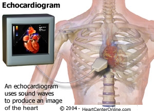
Echocardiogram
An echocardiogram is a test in which ultrasound is used to examine the heart.
Echocardiogram
(source: howstuffworks.com)
An Echocardiogram is a painless procedure that uses an ultrasound beam to view the heart in motion. The procedure is similar to that used to monitor a fetus. An ultrasonic transducer, which looks like a microphone, transmits and receives the ultrasound waves. It is placed on the chest wall and moved around to view different heart structures. Ultrasound waves are reflected only when they reach the edge of two structures with different densities. The reflected waves produce a moving image of the edges of heart structures which are displayed on a screen and recorded on tape.
The types of Echocardiograms are M-mode, 2-D and doppler. M-mode is a one dimensional view of a small section of the heart as it moves. 2-D echocardiogram produces a moving two dimensional slice of the heart. Doppler ultrasound is used to evaluate the velocity and turbulence of blood flow in the heart.
Echocardiograms can evaluate:
- the presence of any abnormal fluid collection in the sac around the heart (pericardium).
- the chamber size, thickness of the heart muscle wall and how well it is functioning.
- the function of the heart valves - whether they are obstructing blood flow or leaking.
any abnormal connections between chambers and vessels that may exist in congenital heart disease.
- wall motion abnormalities that occur when the heart muscle is not receiving enough blood.
- the presence of aneurysms, clots, tumors, vegetations (bacterial growths) on the valves.
Echo Test
Source: heartsite.com
Sticky patches or electrodes are attached to the chest and shoulders and connected to electrodes or wires. These help to record the electrocardiogram (EKG or ECG) during the echocardiography test. The EKG helps in the timing of various cardiac events (filling and emptying of chambers). A colorless gel is then applied to the chest and the echo transducer is placed on top of it. The echo technologist then makes recordings from different parts of the chest to obtain several views of the heart. You may be asked to move form your back and to the side. Instructions may also be given for you to breathe slowly or to hold your breath. This helps in obtaining higher quality pictures. The images are constantly viewed on the monitor. It is also recorded on photographic paper and on videotape. The tape offers a permanent record of the examination and is reviewed by the physician prior to completion of the final report.
Size of the chambers of the heart, including the dimension or volume of the cavity and the thickness of the walls. The appearance of the walls may also help identify certain types of heart disease that predominantly involve the heart muscle. In patients with long standing hypertension or high blood pressure, the test can determine the thickness and "stiffness" of the LV walls. When the LV pump function is reduced in patients with heart failure, the LV and RV tends to dilate or enlarge. Echocardiography can measure the severity of this enlargement. Serial studies performed on an annual basis can gauge the response of treatment.
Pumping function of the heart can be assessed by echocardiography. One can tell if the pumping power of the heart is normal or reduced to a mild or severe degree. This measure is known as an ejection fraction or EF. A normal EF is around 55 to 65%. Numbers below 45% usually represent some decrease in the pumping strength of the heart, while numbers below 30 to 35% are representative of an important decrease.
How safe is echocardiography?
Echocardiography is extremely safe. There are no known risks from the clinical use of ultrasound during this type of testing.
How long does it take?
A brief examination in an uncomplicated case may be done within 15 to 20 minutes. The additional use of Doppler may add an additional 10 to 20 minutes. However, it may take up to an hour when there are multiple problems or when there are technical problems (for example, patients with lung disease, obesity, restlessness, and significant shortness of breath may be more difficult to image).
|